
Page 507 - Atlas of Histology with Functional Correlations
P. 507
TONGUE
The tongue is a muscular organ located in the oral cavity (Fig. 13.2). The core of
the tongue consists of connective tissue and interlacing bundles of skeletal
muscle fibers. The distribution and random orientation of individual skeletal
muscle fibers in the tongue allows for its increased movement during chewing,
swallowing, and speaking. The dorsal surface of the tongue is divided into an
anterior two-thirds and a posterior one-third section by a V-shaped depression
called the sulcus terminalis.
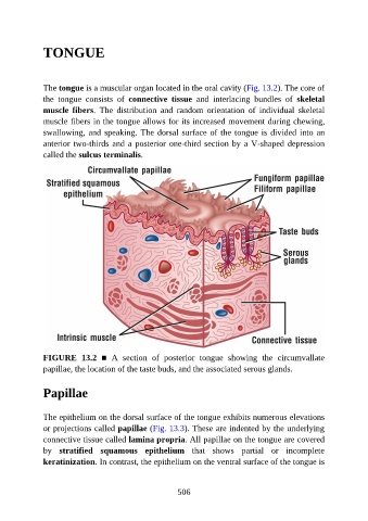
FIGURE 13.2 ■ A section of posterior tongue showing the circumvallate
papillae, the location of the taste buds, and the associated serous glands.
Papillae
The epithelium on the dorsal surface of the tongue exhibits numerous elevations
or projections called papillae (Fig. 13.3). These are indented by the underlying
connective tissue called lamina propria. All papillae on the tongue are covered
by stratified squamous epithelium that shows partial or incomplete
keratinization. In contrast, the epithelium on the ventral surface of the tongue is
506