
Page 511 - Atlas of Histology with Functional Correlations
P. 511
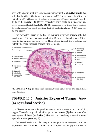
lined with a moist, stratified, squamous nonkeratinized oral epithelium (8) that

               is thicker than the epithelium of the epidermis (11). The surface cells of the oral
               epithelium  (8),  without  cornification,  are  sloughed  off  (desquamated)  into  the
               fluids  of  the  mouth  (10).  Deeper  connective  tissue  contains  tubuloacinar  and
               mucus-secreting labial glands (9, 18). The secretions from these glands moisten
               the oral mucosa. The small excretory ducts of the labial glands (9, 18) open into

               the oral cavity.

                   The connective tissue of the lip also contains numerous adipose cells (7),
               blood vessels (6), and numerous capillaries. Because the blood vessels (6) are
               close  to  the  surface,  the  color  of  the  blood  shows  through  the  overlying  thin

               epithelium, giving the lips a characteristic red color.


               FIGURE 13.5 ■ Lip (longitudinal section). Stain: hematoxylin and eosin. Low
               magnification.


               FIGURE 13.6 | Anterior Region of Tongue: Apex

               (Longitudinal Section)


               This  illustration  shows  a  longitudinal  section  of  the  anterior  portion  of  the
               tongue. The oral cavity is lined with a protective mucosa (5) that consists of an

               outer  epithelial  layer  (epithelium)  (5a)  and  an  underlying  connective  tissue
               layer, the lamina propria (5b).

                   The  dorsal  surface  of  the  tongue  is  rough  due  to  numerous  mucosal
               projections called papillae (1, 2, 6). In contrast, the mucosa (5) of the ventral


                                                          510