
Page 515 - Atlas of Histology with Functional Correlations
P. 515
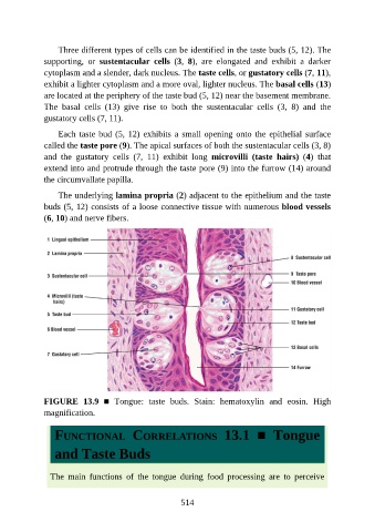
Three different types of cells can be identified in the taste buds (5, 12). The

               supporting,  or  sustentacular  cells  (3,  8),  are  elongated  and  exhibit  a  darker
               cytoplasm and a slender, dark nucleus. The taste cells, or gustatory cells (7, 11),
               exhibit a lighter cytoplasm and a more oval, lighter nucleus. The basal cells (13)
               are located at the periphery of the taste bud (5, 12) near the basement membrane.
               The  basal  cells  (13)  give  rise  to  both  the  sustentacular  cells  (3,  8)  and  the

               gustatory cells (7, 11).

                   Each taste bud (5, 12) exhibits a small opening onto the epithelial surface
               called the taste pore (9). The apical surfaces of both the sustentacular cells (3, 8)
               and  the  gustatory  cells  (7,  11)  exhibit  long  microvilli  (taste  hairs)  (4)  that

               extend into and protrude through the taste pore (9) into the furrow (14) around
               the circumvallate papilla.

                   The underlying lamina propria (2) adjacent to the epithelium and the taste
               buds (5, 12) consists of a loose connective tissue with numerous blood vessels
               (6, 10) and nerve fibers.


               FIGURE  13.9  ■  Tongue:  taste  buds.  Stain:  hematoxylin  and  eosin.  High
               magnification.


                  FUNCTIONAL  CORRELATIONS  13.1  ■  Tongue


                  and Taste Buds


                 The  main  functions  of  the  tongue  during  food  processing  are  to  perceive


                                                          514