
Page 518 - Atlas of Histology with Functional Correlations
P. 518
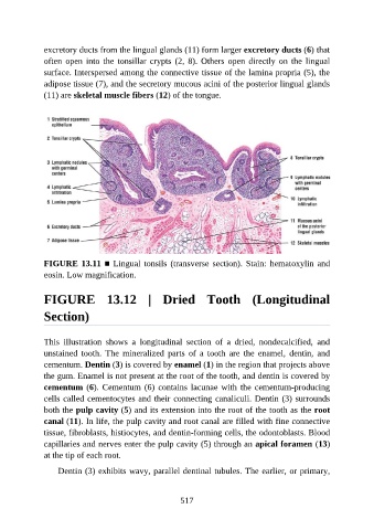
excretory ducts from the lingual glands (11) form larger excretory ducts (6) that

               often  open  into  the  tonsillar  crypts  (2,  8).  Others  open  directly  on  the  lingual
               surface. Interspersed among the connective tissue of the lamina propria (5), the
               adipose tissue (7), and the secretory mucous acini of the posterior lingual glands
               (11) are skeletal muscle fibers (12) of the tongue.


               FIGURE 13.11 ■ Lingual tonsils (transverse section). Stain: hematoxylin and
               eosin. Low magnification.


               FIGURE  13.12  |  Dried  Tooth  (Longitudinal


               Section)


               This  illustration  shows  a  longitudinal  section  of  a  dried,  nondecalcified,  and

               unstained  tooth.  The  mineralized  parts  of  a  tooth  are  the  enamel,  dentin,  and
               cementum. Dentin (3) is covered by enamel (1) in the region that projects above
               the gum. Enamel is not present at the root of the tooth, and dentin is covered by
               cementum (6). Cementum (6) contains lacunae with the cementum-producing
               cells called cementocytes and their connecting canaliculi. Dentin (3) surrounds

               both the pulp cavity (5) and its extension into the root of the tooth as the root
               canal (11). In life, the pulp cavity and root canal are filled with fine connective
               tissue, fibroblasts, histiocytes, and dentin-forming cells, the odontoblasts. Blood

               capillaries and nerves enter the pulp cavity (5) through an apical foramen (13)
               at the tip of each root.

                   Dentin (3) exhibits wavy, parallel dentinal tubules. The earlier, or primary,


                                                          517