
Page 517 - Atlas of Histology with Functional Correlations
P. 517
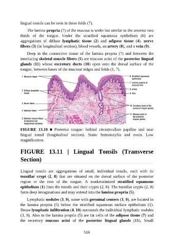
lingual tonsils can be seen in these folds (7).

                   The lamina propria (7) of the mucosa is wider but similar to the anterior two

               thirds  of  the  tongue.  Under  the  stratified  squamous  epithelium  (6)  are
               aggregations  of  diffuse  lymphatic  tissue  (2)  and  adipose  tissue  (4),  nerve
               fibers (3) (in longitudinal section), blood vessels, an artery (8), and a vein (9).

                   Deep  in  the  connective  tissue  of  the  lamina  propria  (7)  and  between  the
               interlacing skeletal muscle fibers (5) are mucous acini of the posterior lingual

               glands  (11)  whose  excretory  ducts  (10)  open  onto  the  dorsal  surface  of  the
               tongue, between bases of the mucosal ridges and folds (1, 7).


               FIGURE  13.10  ■  Posterior  tongue:  behind  circumvallate  papillae  and  near
               lingual  tonsil  (longitudinal  section).  Stain:  hematoxylin  and  eosin.  Low
               magnification.


               FIGURE  13.11  |  Lingual  Tonsils  (Transverse

               Section)


               Lingual  tonsils  are  aggregations  of  small,  individual  tonsils,  each  with  its

               tonsillar  crypt  (2,  8)  that  are  situated  on  the  dorsal  surface  of  the  posterior
               region  or  the  root  of  the  tongue.  A  nonkeratinized  stratified  squamous
               epithelium (1) lines the tonsils and their crypts (2, 8). The tonsillar crypts (2, 8)

               form deep invaginations and may extend into the lamina propria (5).

                   Lymphatic nodules (3, 9), some with germinal centers (3, 9), are located in
               the  lamina  propria  (5)  below  the  stratified  squamous  surface  epithelium  (1).
               Dense lymphatic infiltration (4, 10) surrounds the individual lymphatic nodules
               (3, 9). Also in the lamina propria (5) are fat cells of the adipose tissue (7) and

               the  secretory  mucous  acini  of  the  posterior  lingual  glands  (11).  Small


                                                          516