
Page 521 - Atlas of Histology with Functional Correlations
P. 521
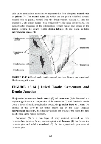
cells called ameloblasts as successive segments that form elongated enamel rods

               or  prisms  (7).  The  enamel  tufts  (6),  which  are  the  poorly  calcified,  twisted
               enamel  rods  or  prisms,  extend  from  the  dentinoenamel  junction  (1)  into  the
               enamel (5). The dentin matrix (4) is produced by cells called odontoblasts. The
               odontoblastic  processes  of  the  odontoblasts  occupy  tunnel-like  spaces  in  the
               dentin,  forming  the  clearly  visible  dentin  tubules  (3)  and  black,  air-filled

               interglobular spaces (2).


               FIGURE 13.13 ■ Dried tooth: dentinoenamel junction. Ground and unstained.
               Medium magnification.


               FIGURE  13.14  |  Dried  Tooth:  Cementum  and

               Dentin Junction


               The junction between the dentin matrix (5) and cementum (2) is illustrated at a

               higher magnification. At the junction of the cementum (2) with the dentin matrix
               (5) is a layer of small interglobular spaces, the granular layer of Tomes (7).
               Internal  to  this  layer  in  the  dentin  matrix  (5)  are  the  large,  irregular
               interglobular spaces (4, 8) commonly seen in the crown of the tooth, but may
               also be seen in the root of the tooth.


                   Cementum  (2)  is  a  thin  layer  of  bony  material  secreted  by  cells
               cementoblasts  (mature  forms,  cementocytes)  with  lacunae  (1)  that  house  the
               cementocytes  and  exhibit  canaliculi  (3)  for  the  cytoplasmic  processes  of
               cementocytes.


                                                          520