
Page 522 - Atlas of Histology with Functional Correlations
P. 522
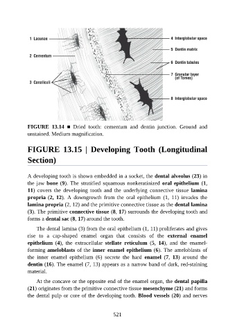
FIGURE  13.14  ■  Dried  tooth:  cementum  and  dentin  junction.  Ground  and

               unstained. Medium magnification.


               FIGURE  13.15  |  Developing  Tooth  (Longitudinal


               Section)


               A developing tooth is shown embedded in a socket, the dental alveolus (23) in
               the jaw bone (9). The  stratified  squamous  nonkeratinized  oral epithelium  (1,

               11)  covers  the  developing  tooth  and  the  underlying  connective  tissue  lamina
               propria (2,  12).  A  downgrowth  from  the  oral  epithelium  (1,  11)  invades  the
               lamina propria (2, 12) and the primitive connective tissue as the dental lamina
               (3). The primitive connective tissue (8, 17) surrounds the developing tooth and
               forms a dental sac (8, 17) around the tooth.


                   The dental lamina (3) from the oral epithelium (1, 11) proliferates and gives
               rise  to  a  cap-shaped  enamel  organ  that  consists  of  the  external  enamel
               epithelium  (4),  the  extracellular  stellate  reticulum  (5,  14),  and  the  enamel-
               forming ameloblasts of the inner enamel epithelium (6). The ameloblasts of

               the  inner  enamel  epithelium  (6)  secrete  the  hard  enamel  (7,  13)  around  the
               dentin (16). The enamel (7, 13) appears as a narrow band of dark, red-staining
               material.

                   At the concave or the opposite end of the enamel organ, the dental papilla
               (21) originates from the primitive connective tissue mesenchyme (21) and forms

               the dental pulp or core of the developing tooth. Blood vessels (20) and nerves


                                                          521