
Page 514 - Atlas of Histology with Functional Correlations
P. 514
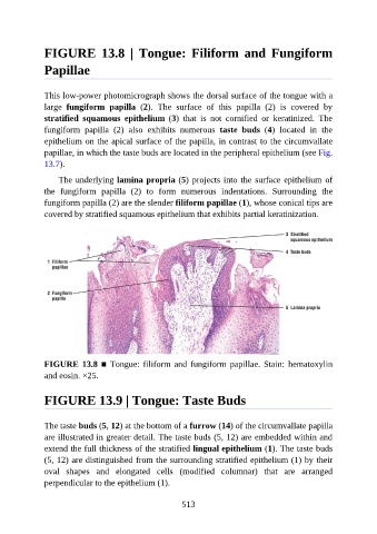
FIGURE  13.8  |  Tongue:  Filiform  and  Fungiform

               Papillae


               This low-power photomicrograph shows the dorsal surface of the tongue with a
               large  fungiform  papilla  (2).  The  surface  of  this  papilla  (2)  is  covered  by

               stratified  squamous  epithelium  (3)  that  is  not  cornified  or  keratinized.  The
               fungiform  papilla  (2)  also  exhibits  numerous  taste  buds  (4)  located  in  the
               epithelium on the apical surface of the papilla, in contrast to the circumvallate
               papillae, in which the taste buds are located in the peripheral epithelium (see Fig.

               13.7).

                   The underlying lamina propria (5) projects into the surface epithelium of
               the  fungiform  papilla  (2)  to  form  numerous  indentations.  Surrounding  the
               fungiform papilla (2) are the slender filiform papillae (1), whose conical tips are

               covered by stratified squamous epithelium that exhibits partial keratinization.


               FIGURE 13.8 ■ Tongue: filiform and fungiform papillae. Stain:  hematoxylin
               and eosin. ×25.


               FIGURE 13.9 | Tongue: Taste Buds


               The taste buds (5, 12) at the bottom of a furrow (14) of the circumvallate papilla

               are illustrated in greater detail. The taste buds (5, 12) are embedded within and
               extend the full thickness of the stratified lingual epithelium (1). The taste buds
               (5, 12) are distinguished from the surrounding stratified epithelium (1) by their
               oval  shapes  and  elongated  cells  (modified  columnar)  that  are  arranged
               perpendicular to the epithelium (1).


                                                          513