
Page 512 - Atlas of Histology with Functional Correlations
P. 512
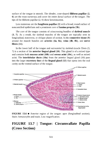
surface of the tongue is smooth. The slender, cone-shaped filiform papillae (2,

               6) are the most numerous and cover the entire dorsal surface of the tongue. The
               tips of the filiform papillae (2, 6) show keratinization.

                   Less numerous are the fungiform papillae (1) with a broad, round surface of
               noncornified epithelium and a prominent core of lamina propria (5b).

                   The core of the tongue consists of crisscrossing bundles of skeletal muscle
               (3,  7).  As  a  result,  the  skeletal  muscles  of  the  tongue  are  typically  seen  in

               longitudinal, transverse, or oblique planes of section. In the connective tissue (9)
               around  the  muscle  bundles  are  arteries  (4a,  8a),  veins  (4b,  8b),  and  nerve
               fibers (11).

                   In the lower half of the tongue and surrounded by skeletal muscle fibers (3,

               7) is a section of the anterior lingual gland (10). This gland is of a mixed type
               and contains both mucous acini (10b) and serous acini (10c), as well as mixed
               acini.  The  interlobular ducts (10a)  from  the  anterior  lingual  gland  (10)  pass
               into the larger excretory duct of the lingual gland (12) that opens into the oral

               cavity on the ventral surface of the tongue.


               FIGURE  13.6  ■  Anterior  region  of  the  tongue:  apex  (longitudinal  section).
               Stain: hematoxylin and eosin. Low magnification.


               FIGURE  13.7  |  Tongue:  Circumvallate  Papilla

               (Cross Section)


                                                          511