
Page 513 - Atlas of Histology with Functional Correlations
P. 513
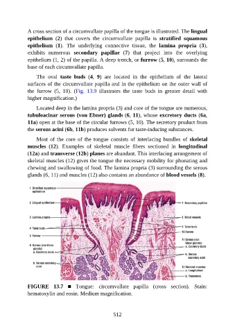
A cross section of a circumvallate papilla of the tongue is illustrated. The lingual

               epithelium  (2)  that  covers  the  circumvallate  papilla  is  stratified  squamous
               epithelium  (1).  The  underlying  connective  tissue,  the  lamina  propria  (3),
               exhibits  numerous  secondary  papillae  (7)  that  project  into  the  overlying
               epithelium (1, 2) of the papilla. A deep trench, or furrow (5, 10), surrounds the
               base of each circumvallate papilla.


                   The  oval  taste  buds  (4,  9)  are  located  in  the  epithelium  of  the  lateral
               surfaces of the circumvallate papilla and in the epithelium on the outer wall of
               the  furrow  (5,  10).  (Fig.  13.9  illustrates  the  taste  buds  in  greater  detail  with
               higher magnification.)


                   Located deep in the lamina propria (3) and core of the tongue are numerous,
               tubuloacinar serous (von Ebner) glands (6, 11), whose excretory ducts (6a,
               11a) open at the base of the circular furrows (5, 10). The secretory product from
               the serous acini (6b, 11b) produces solvents for taste-inducing substances.

                   Most  of  the  core  of  the  tongue  consists  of  interlacing  bundles  of  skeletal

               muscles  (12).  Examples  of  skeletal  muscle  fibers  sectioned  in  longitudinal
               (12a) and transverse (12b) planes are abundant. This interlacing arrangement of
               skeletal muscles (12) gives the tongue the necessary mobility for phonating and
               chewing and swallowing of food. The lamina propria (3) surrounding the serous

               glands (6, 11) and muscles (12) also contains an abundance of blood vessels (8).


               FIGURE  13.7  ■  Tongue:  circumvallate  papilla  (cross  section).  Stain:
               hematoxylin and eosin. Medium magnification.


                                                          512