
Page 545 - Atlas of Histology with Functional Correlations
P. 545
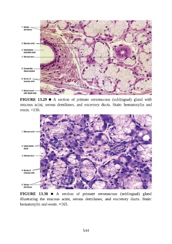
FIGURE  13.29  ■  A  section  of  primate  seromucous  (sublingual)  gland  with
               mucous  acini,  serous  demilunes,  and  excretory  ducts.  Stain:  hematoxylin  and

               eosin. ×130.


               FIGURE  13.30  ■  A  section  of  primate  seromucous  (sublingual)  gland
               illustrating  the  mucous  acini,  serous  demilunes,  and  excretory  ducts.  Stain:
               hematoxylin and eosin. ×165.


                                                          544