
Page 544 - Atlas of Histology with Functional Correlations
P. 544
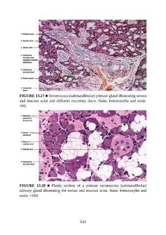
FIGURE 13.27 ■ Seromucous (submandibular) primate gland illustrating serous
               and mucous acini and different excretory ducts. Stain: hematoxylin and eosin.

               ×60.


               FIGURE  13.28  ■  Plastic  section  of  a  primate  seromucous  (submandibular)

               salivary gland illustrating the serous and mucous acini. Stain: hematoxylin and
               eosin. ×160.


                                                          543