
Page 588 - Atlas of Histology with Functional Correlations
P. 588
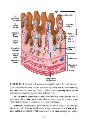
FIGURE 15.1 ■ Different cell types and layers in the wall of the small intestine.

               Each villus contains blood vessels, lymphatic capillaries, nerves, smooth muscle,
               and loose irregular connective tissue, in addition to the lamina propria plasma
               cells, tissue eosinophils, macrophages, and mast cells.

                   Smooth muscle fibers from the muscularis mucosae extend into the core of

               individual villi to induce movements. This action increases the contacts of the
               villi with the digested food products in the intestinal lumen.

                   Microvilli are cytoplasmic extensions that cover the apices of the intestinal
               absorptive cells. They are visible under a light microscope as a brush border

               (also called striated border). With transmission electron microscopy, they appear


                                                          587