
Page 592 - Atlas of Histology with Functional Correlations
P. 592
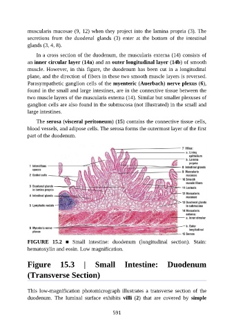
muscularis mucosae (9, 12) when they project into the lamina propria (3). The

               secretions  from  the  duodenal  glands  (3)  enter  at  the  bottom  of  the  intestinal
               glands (3, 4, 8).

                   In a cross section of the duodenum, the muscularis externa (14) consists of
               an inner circular layer (14a) and an outer longitudinal layer (14b) of smooth
               muscle. However, in this figure, the duodenum has been cut in a longitudinal

               plane, and the direction of fibers in these two smooth muscle layers is reversed.
               Parasympathetic ganglion cells of the myenteric (Auerbach) nerve plexus (6),
               found in the small and large intestines, are in the connective tissue between the
               two muscle layers of the muscularis externa (14). Similar but smaller plexuses of

               ganglion cells are also found in the submucosa (not illustrated) in the small and
               large intestines.

                   The serosa (visceral peritoneum) (15) contains the connective tissue cells,
               blood vessels, and adipose cells. The serosa forms the outermost layer of the first
               part of the duodenum.


               FIGURE  15.2  ■  Small  intestine:  duodenum  (longitudinal  section).  Stain:
               hematoxylin and eosin. Low magnification.


               Figure  15.3  |  Small  Intestine:  Duodenum

               (Transverse Section)


               This low-magnification photomicrograph illustrates  a  transverse  section  of  the
               duodenum.  The  luminal  surface  exhibits  villi  (2)  that  are  covered  by  simple


                                                          591