
Page 595 - Atlas of Histology with Functional Correlations
P. 595
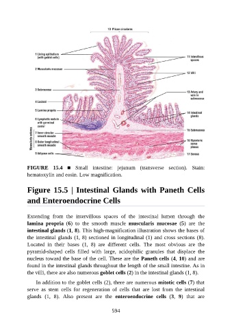
FIGURE  15.4  ■  Small  intestine:  jejunum  (transverse  section).  Stain:
               hematoxylin and eosin. Low magnification.


               Figure 15.5 | Intestinal Glands with Paneth Cells

               and Enteroendocrine Cells


               Extending  from  the  intervillous  spaces  of  the  intestinal  lumen  through  the

               lamina  propria  (6)  to  the  smooth  muscle  muscularis  mucosae  (5)  are  the
               intestinal glands (1, 8). This high-magnification illustration shows the bases of
               the intestinal glands (1, 8) sectioned in longitudinal (1) and cross sections (8).

               Located  in  their  bases  (1,  8)  are  different  cells.  The  most  obvious  are  the
               pyramid-shaped  cells  filled  with  large,  acidophilic  granules  that  displace  the
               nucleus toward the base of the cell. These are the Paneth cells (4, 10) and are
               found in the intestinal glands throughout the length of the small intestine. As in
               the villi, there are also numerous goblet cells (2) in the intestinal glands (1, 8).


                   In addition to the goblet cells (2), there are numerous mitotic cells (7) that
               serve  as  stem  cells  for  regeneration  of  cells  that  are  lost  from  the  intestinal
               glands  (1,  8).  Also  present  are  the  enteroendocrine  cells  (3,  9)  that  are


                                                          594