
Page 600 - Atlas of Histology with Functional Correlations
P. 600
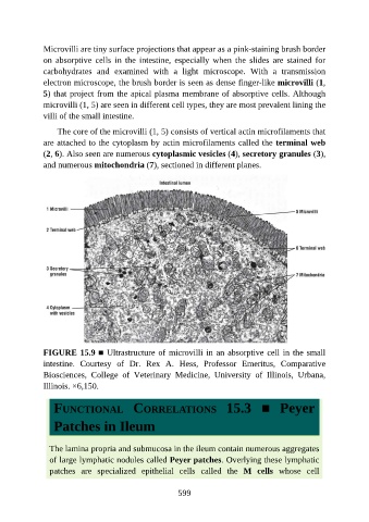
Microvilli are tiny surface projections that appear as a pink-staining brush border

               on  absorptive  cells  in  the  intestine,  especially  when  the  slides  are  stained  for
               carbohydrates  and  examined  with  a  light  microscope.  With  a  transmission
               electron microscope, the brush border is seen as dense finger-like microvilli (1,
               5) that project from the apical plasma membrane of absorptive cells. Although
               microvilli (1, 5) are seen in different cell types, they are most prevalent lining the

               villi of the small intestine.

                   The core of the microvilli (1, 5) consists of vertical actin microfilaments that
               are attached to the cytoplasm by actin microfilaments called the terminal web
               (2, 6). Also seen are numerous cytoplasmic vesicles (4), secretory granules (3),

               and numerous mitochondria (7), sectioned in different planes.


               FIGURE 15.9 ■ Ultrastructure of microvilli in an absorptive cell in the small

               intestine.  Courtesy  of  Dr.  Rex  A.  Hess,  Professor  Emeritus,  Comparative
               Biosciences,  College  of  Veterinary  Medicine,  University  of  Illinois,  Urbana,
               Illinois. ×6,150.


                  FUNCTIONAL  CORRELATIONS  15.3  ■  Peyer


                  Patches in Ileum


                 The lamina propria and submucosa in the ileum contain numerous aggregates
                 of large lymphatic nodules called Peyer patches. Overlying these lymphatic
                 patches  are  specialized  epithelial  cells  called  the  M  cells  whose  cell


                                                          599