
Page 605 - Atlas of Histology with Functional Correlations
P. 605
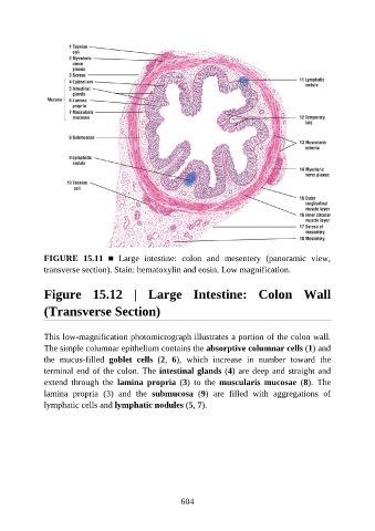
FIGURE  15.11  ■  Large  intestine:  colon  and  mesentery  (panoramic  view,

               transverse section). Stain: hematoxylin and eosin. Low magnification.


               Figure  15.12  |  Large  Intestine:  Colon  Wall


               (Transverse Section)


               This low-magnification photomicrograph illustrates a portion of the colon wall.
               The simple columnar epithelium contains the absorptive columnar cells (1) and

               the  mucus-filled  goblet  cells  (2,  6),  which  increase  in  number  toward  the
               terminal end of the colon. The intestinal glands (4) are deep and straight and
               extend  through  the  lamina  propria  (3)  to  the  muscularis  mucosae  (8).  The
               lamina  propria  (3)  and  the  submucosa  (9)  are  filled  with  aggregations  of

               lymphatic cells and lymphatic nodules (5, 7).


                                                          604