
Page 607 - Atlas of Histology with Functional Correlations
P. 607
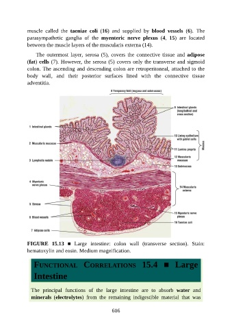
muscle  called  the  taeniae  coli  (16)  and  supplied  by  blood  vessels  (6).  The

               parasympathetic  ganglia  of  the  myenteric  nerve  plexus  (4,  15)  are  located
               between the muscle layers of the muscularis externa (14).

                   The  outermost  layer,  serosa  (5),  covers  the  connective  tissue  and  adipose
               (fat) cells (7). However, the serosa (5) covers only the transverse and sigmoid
               colon. The ascending and descending colon are retroperitoneal, attached to the

               body  wall,  and  their  posterior  surfaces  lined  with  the  connective  tissue
               adventitia.


               FIGURE  15.13  ■  Large  intestine:  colon  wall  (transverse  section).  Stain:

               hematoxylin and eosin. Medium magnification.


                  FUNCTIONAL  CORRELATIONS  15.4  ■  Large

                  Intestine


                 The  principal  functions  of  the  large  intestine  are  to  absorb  water  and
                 minerals  (electrolytes)  from  the  remaining  indigestible  material  that  was


                                                          606