
Page 610 - Atlas of Histology with Functional Correlations
P. 610
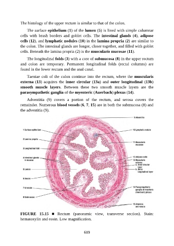
The histology of the upper rectum is similar to that of the colon.

                   The surface epithelium (1) of the lumen (5) is lined with simple columnar

               cells  with  brush  borders  and  goblet  cells.  The  intestinal  glands  (4),  adipose
               cells (12), and lymphatic nodules (10) in the lamina propria (2) are similar to
               the colon. The intestinal glands are longer, closer together, and filled with goblet
               cells. Beneath the lamina propria (2) is the muscularis mucosae (11).

                   The longitudinal folds (3) with a core of submucosa (8) in the upper rectum

               and  colon  are  temporary.  Permanent  longitudinal  folds  (rectal  columns)  are
               found in the lower rectum and the anal canal.

                   Taeniae coli of the colon continue into the rectum, where the muscularis
               externa (13)  acquires  the  inner circular (13a)  and  outer  longitudinal  (13b)

               smooth  muscle  layers.  Between  these  two  smooth  muscle  layers  are  the
               parasympathetic ganglia of the myenteric (Auerbach) plexus (14).

                   Adventitia  (9)  covers  a  portion  of  the  rectum,  and  serosa  covers  the
               remainder. Numerous blood vessels (6, 7, 15) are in both the submucosa (8) and
               the adventitia (9).


               FIGURE  15.15  ■  Rectum  (panoramic  view,  transverse  section).  Stain:
               hematoxylin and eosin. Low magnification.


                                                          609