
Page 611 - Atlas of Histology with Functional Correlations
P. 611
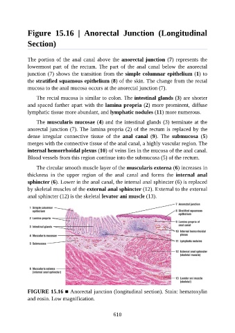
Figure  15.16  |  Anorectal  Junction  (Longitudinal

               Section)


               The portion of the anal canal above the anorectal junction (7) represents the
               lowermost  part  of  the  rectum.  The  part  of  the  anal  canal  below  the  anorectal

               junction (7) shows the transition from the simple columnar epithelium (1) to
               the stratified squamous epithelium (8) of the skin. The change from the rectal
               mucosa to the anal mucosa occurs at the anorectal junction (7).

                   The rectal mucosa is similar to colon. The intestinal glands (3) are shorter
               and spaced farther apart with the lamina propria (2) more prominent, diffuse

               lymphatic tissue more abundant, and lymphatic nodules (11) more numerous.

                   The muscularis mucosae (4) and the intestinal glands (3) terminate at the
               anorectal junction (7). The lamina propria (2) of the rectum is replaced by the
               dense  irregular  connective  tissue  of  the  anal  canal  (9).  The  submucosa  (5)

               merges with the connective tissue of the anal canal, a highly vascular region. The
               internal hemorrhoidal plexus (10) of veins lies in the mucosa of the anal canal.
               Blood vessels from this region continue into the submucosa (5) of the rectum.

                   The circular smooth muscle layer of the muscularis externa (6) increases in

               thickness  in  the  upper  region  of  the  anal  canal  and  forms  the  internal  anal
               sphincter (6). Lower in the anal canal, the internal anal sphincter (6) is replaced
               by skeletal muscles of the external anal sphincter (12). External to the external
               anal sphincter (12) is the skeletal levator ani muscle (13).


               FIGURE 15.16 ■ Anorectal junction (longitudinal section). Stain: hematoxylin
               and eosin. Low magnification.


                                                          610