
Page 606 - Atlas of Histology with Functional Correlations
P. 606
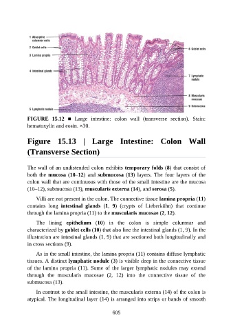
FIGURE  15.12  ■  Large  intestine:  colon  wall  (transverse  section).  Stain:
               hematoxylin and eosin. ×30.


               Figure  15.13  |  Large  Intestine:  Colon  Wall

               (Transverse Section)


               The wall of an undistended colon exhibits temporary folds (8) that consist of
               both  the  mucosa (10–12)  and  submucosa (13)  layers.  The  four  layers  of  the
               colon wall that are continuous with those of the small intestine are the mucosa
               (10–12), submucosa (13), muscularis externa (14), and serosa (5).

                   Villi are not present in the colon. The connective tissue lamina propria (11)

               contains  long  intestinal  glands  (1,  9)  (crypts  of  Lieberkühn)  that  continue
               through the lamina propria (11) to the muscularis mucosae (2, 12).

                   The  lining  epithelium  (10)  in  the  colon  is  simple  columnar  and
               characterized by goblet cells (10) that also line the intestinal glands (1, 9). In the

               illustration are intestinal glands (1, 9) that are sectioned both longitudinally and
               in cross sections (9).

                   As in the small intestine, the lamina propria (11) contains diffuse lymphatic
               tissues. A distinct lymphatic nodule (3) is visible deep in the connective tissue
               of the lamina propria (11). Some of the larger lymphatic nodules may extend

               through  the  muscularis  mucosae  (2,  12)  into  the  connective  tissue  of  the
               submucosa (13).

                   In contrast to the small intestine, the muscularis externa (14) of the colon is
               atypical. The longitudinal layer (14) is arranged into strips or bands of smooth


                                                          605