
Page 609 - Atlas of Histology with Functional Correlations
P. 609
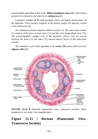
spaced farther apart than in the colon. Diffuse lymphatic tissue (6) in the lamina

               propria (3) is abundant and often in the submucosa (8).

                   Lymphatic nodules (4, 9) with germinal centers are highly characteristic of
               the appendix. These nodules originate in the lamina propria (3) and may extend
               to the submucosa (8).

                   The submucosa (8) has numerous blood vessels (11). The muscularis externa
               (7) consists of the inner circular layer (7a) and the outer longitudinal layer (7b).

               The  parasympathetic  ganglia  (12)  of  the  myenteric  plexus  (12)  are  located
               between the inner (7a) and outer (7b) smooth muscle layers of the muscularis
               externa.

                   The outermost layer of the appendix is the serosa (10) under which are seen

               adipose cells (13).


               FIGURE  15.14  ■  Appendix  (panoramic  view,  transverse  section).  Stain:
               hematoxylin and eosin. Low magnification.


               Figure  15.15  |  Rectum  (Panoramic  View,

               Transverse Section)


                                                          608