
Page 599 - Atlas of Histology with Functional Correlations
P. 599
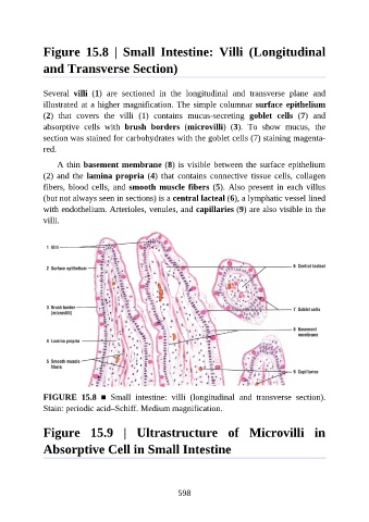
Figure  15.8  |  Small  Intestine:  Villi  (Longitudinal

               and Transverse Section)


               Several  villi  (1)  are  sectioned  in  the  longitudinal  and  transverse  plane  and
               illustrated at a higher magnification. The simple columnar surface epithelium

               (2)  that  covers  the  villi  (1)  contains  mucus-secreting  goblet  cells  (7)  and
               absorptive  cells  with  brush  borders  (microvilli)  (3).  To  show  mucus,  the
               section was stained for carbohydrates with the goblet cells (7) staining magenta-
               red.

                   A thin basement membrane (8) is visible between the surface epithelium

               (2) and the lamina propria (4)  that  contains  connective  tissue  cells,  collagen
               fibers, blood cells, and smooth muscle fibers (5). Also present in each villus
               (but not always seen in sections) is a central lacteal (6), a lymphatic vessel lined

               with endothelium. Arterioles, venules, and capillaries (9) are also visible in the
               villi.


               FIGURE  15.8  ■  Small  intestine:  villi  (longitudinal  and  transverse  section).

               Stain: periodic acid–Schiff. Medium magnification.


               Figure  15.9  |  Ultrastructure  of  Microvilli  in


               Absorptive Cell in Small Intestine


                                                          598