
Page 594 - Atlas of Histology with Functional Correlations
P. 594
bicarbonate secretions from the duodenal glands buffer or neutralize the
acidic chyme. This action provides a more favorable environment for
digestive enzymes that are released into the duodenum from the pancreas.
Enteroendocrine cells located in the secretory acini of duodenal
(Brunner) glands also produce a polypeptide hormone called urogastrone
that inhibits or decreases hydrochloric acid secretion by the parietal cells in
the stomach.
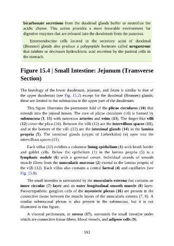
Figure 15.4 | Small Intestine: Jejunum (Transverse
Section)
The histology of the lower duodenum, jejunum, and ileum is similar to that of
the upper duodenum (see Fig. 15.2) except for the duodenal (Brunner) glands;
these are limited to the submucosa in the upper part of the duodenum.
This figure illustrates the permanent fold of the plicae circulares (10) that
extends into the jejunal lumen. The core of plicae circulares (10) is formed by
submucosa (3, 15) with numerous arteries and veins (13). The finger-like villi
(12) cover the plica (10). Between the villi (12) are the intervillous spaces (11),
and at the bottom of the villi (12) are the intestinal glands (14) in the lamina
propria (5). The intestinal glands (crypts of Lieberkühn) (4) open into the
intervillous spaces (11).
Each villus (12) exhibits a columnar lining epithelium (1) with brush border
and goblet cells. Below the epithelium (1) in the lamina propria (5) is a
lymphatic nodule (6) with a germinal center. Individual strands of smooth
muscle fibers from the muscularis mucosae (2) extend in the lamina propria of
the villi (12). Each villus also contains a central lacteal (4) and capillaries (see
Fig. 15.8).
The small intestine is surrounded by the muscularis externa that contains an
inner circular (7) layer and an outer longitudinal smooth muscle (8) layer.
Parasympathetic ganglion cells of the myenteric plexus (16) are present in the
connective tissue between the muscle layers of the muscularis externa (7, 8). A
similar submucosal plexus is also present in the submucosa, but it is not
illustrated in this figure.
A visceral peritoneum, or serosa (17), surrounds the small intestine under
which are connective tissue fibers, blood vessels, and adipose cells (9).
593