
Page 598 - Atlas of Histology with Functional Correlations
P. 598
Cholecystokinin increases the secretion of pancreatic enzymes into the small
intestine and induces gallbladder contractions to expel the stored bile.
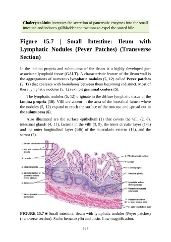
Figure 15.7 | Small Intestine: Ileum with
Lymphatic Nodules (Peyer Patches) (Transverse
Section)
In the lamina propria and submucosa of the ileum is a highly developed gut-
associated lymphoid tissue (GALT). A characteristic feature of the ileum wall is
the aggregations of numerous lymphatic nodules (5, 12) called Peyer patches
(5, 12) that coalesce with boundaries between them becoming indistinct. Most of
these lymphatic nodules (5, 12) exhibit germinal centers (5).
The lymphatic nodules (5, 12) originate in the diffuse lymphatic tissue of the
lamina propria (10). Villi are absent in the area of the intestinal lumen where
the nodules (5, 12) expand to reach the surface of the mucosa and spread out in
the submucosa (6).
Also illustrated are the surface epithelium (1) that covers the villi (2, 8),
intestinal glands (4, 11), lacteals in the villi (3, 9), the inner circular layer (14a)
and the outer longitudinal layer (14b) of the muscularis externa (14), and the
serosa (7).
FIGURE 15.7 ■ Small intestine: ileum with lymphatic nodules (Peyer patches)
(transverse section). Stain: hematoxylin and eosin. Low magnification.
597