
Page 596 - Atlas of Histology with Functional Correlations
P. 596
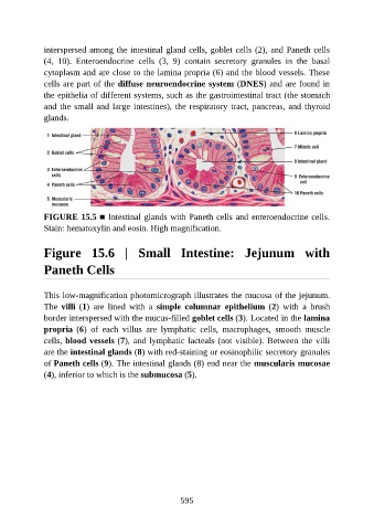
interspersed among the intestinal gland cells, goblet cells (2), and Paneth cells

               (4,  10).  Enteroendocrine  cells  (3,  9)  contain  secretory  granules  in  the  basal
               cytoplasm and are close to the lamina propria (6) and the blood vessels. These
               cells are part of the diffuse neuroendocrine system (DNES) and are found in
               the epithelia of different systems, such as the gastrointestinal tract (the stomach
               and the small and large intestines), the respiratory tract, pancreas, and thyroid

               glands.


               FIGURE 15.5 ■ Intestinal glands with Paneth cells and enteroendocrine cells.

               Stain: hematoxylin and eosin. High magnification.


               Figure  15.6  |  Small  Intestine:  Jejunum  with


               Paneth Cells


               This low-magnification photomicrograph illustrates the mucosa of the jejunum.
               The  villi  (1)  are  lined  with  a  simple  columnar  epithelium  (2)  with  a  brush
               border interspersed with the mucus-filled goblet cells (3). Located in the lamina

               propria  (6)  of  each  villus  are  lymphatic  cells,  macrophages,  smooth  muscle
               cells, blood vessels (7), and lymphatic lacteals (not visible). Between the villi
               are the intestinal glands (8) with red-staining or eosinophilic secretory granules

               of Paneth cells (9). The intestinal glands (8) end near the muscularis mucosae
               (4), inferior to which is the submucosa (5).


                                                          595