Page 593 - Atlas of Histology with Functional Correlations
P. 593
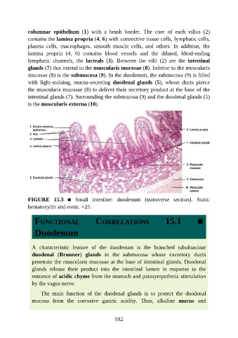
columnar  epithelium  (1)  with  a  brush  border.  The  core  of  each  villus  (2)

               contains the lamina propria (4, 6) with connective tissue cells, lymphatic cells,
               plasma  cells,  macrophages,  smooth  muscle  cells,  and  others.  In  addition,  the
               lamina  propria  (4,  6)  contains  blood  vessels  and  the  dilated,  blind-ending
               lymphatic  channels,  the  lacteals  (3).  Between  the  villi  (2)  are  the  intestinal
               glands (7) that extend to the muscularis mucosae (8). Inferior to the muscularis

               mucosae (8) is the submucosa (9). In the duodenum, the submucosa (9) is filled
               with  light-staining,  mucus-secreting  duodenal glands  (5),  whose  ducts  pierce
               the muscularis mucosae (8) to deliver their secretory product at the base of the

               intestinal glands (7). Surrounding the submucosa (9) and the duodenal glands (5)
               is the muscularis externa (10).


               FIGURE  15.3  ■  Small  intestine:  duodenum  (transverse  section).  Stain:
               hematoxylin and eosin. ×25.


                  FUNCTIONAL                     CORRELATIONS                       15.1            ■


                  Duodenum


                 A  characteristic  feature  of  the  duodenum  is  the  branched  tubuloacinar

                 duodenal  (Brunner)  glands  in  the  submucosa  whose  excretory  ducts
                 penetrate the muscularis mucosae at the base of intestinal glands. Duodenal
                 glands  release  their  product  into  the  intestinal  lumen  in  response  to  the
                 entrance of acidic chyme from the stomach and parasympathetic stimulation

                 by the vagus nerve.

                     The  main  function  of  the  duodenal  glands  is  to  protect  the  duodenal
                 mucosa  from  the  corrosive  gastric  acidity.  Thus,  alkaline  mucus  and


                                                          592

