
Page 60 - Atlas of Histology with Functional Correlations
P. 60
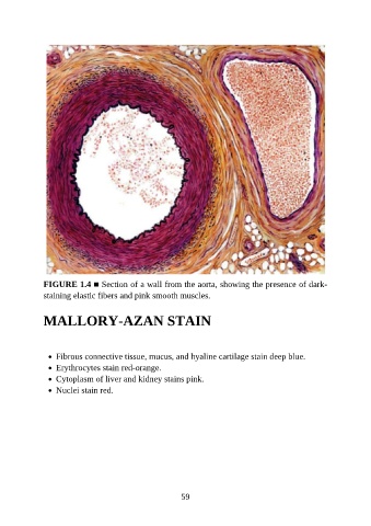
FIGURE 1.4 ■ Section of a wall from the aorta, showing the presence of dark-
               staining elastic fibers and pink smooth muscles.


               MALLORY-AZAN STAIN


                   Fibrous connective tissue, mucus, and hyaline cartilage stain deep blue.
                   Erythrocytes stain red-orange.
                   Cytoplasm of liver and kidney stains pink.
                   Nuclei stain red.


                                                           59