
Page 61 - Atlas of Histology with Functional Correlations
P. 61
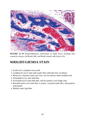
FIGURE  1.5  ■  Intramembranous  ossification  in  skull  bones  showing  blue
               connective tissue, red blood cells, and blood vessels with blood cells.


               WRIGHT/GIEMSA STAIN


                   Erythrocyte cytoplasm stains pink.

                   Lymphocyte nuclei stain dark purple-blue with pale-blue cytoplasm.
                   Monocyte cytoplasm stains pale blue, and the nucleus stains medium blue.
                   Neutrophil nuclei stain dark blue.

                   Eosinophil nuclei stain dark blue, and the granules stain bright pink.
                   Basophil nuclei stain dark blue or purple, cytoplasm pale blue, and granules
                   deep purple.
                   Platelets stain light blue.


                                                           60