
Page 65 - Atlas of Histology with Functional Correlations
P. 65
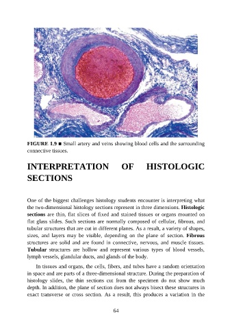
FIGURE 1.9 ■ Small artery and veins showing blood cells and the surrounding

               connective tissues.


               INTERPRETATION                                  OF           HISTOLOGIC


               SECTIONS


               One of the biggest challenges histology students encounter is interpreting what

               the two-dimensional histology sections represent in three dimensions. Histologic
               sections are thin, flat slices of fixed and stained tissues or organs mounted on
               flat glass slides. Such sections are normally composed of cellular, fibrous, and
               tubular structures that are cut in different planes. As a result, a variety of shapes,

               sizes,  and  layers  may  be  visible,  depending  on  the  plane  of  section.  Fibrous
               structures  are  solid  and  are  found  in  connective,  nervous,  and  muscle  tissues.
               Tubular  structures  are  hollow  and  represent  various  types  of  blood  vessels,
               lymph vessels, glandular ducts, and glands of the body.

                   In tissues and organs, the cells, fibers, and tubes have a random orientation

               in space and are parts of a three-dimensional structure. During the preparation of
               histology  slides,  the  thin  sections  cut  from  the  specimen  do  not  show  much
               depth. In addition, the plane of section does not always bisect these structures in

               exact  transverse  or  cross  section.  As  a  result,  this  produces  a  variation  in  the


                                                           64