
Page 704 - Atlas of Histology with Functional Correlations
P. 704
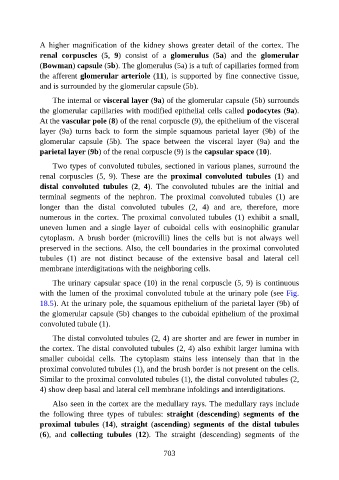
A  higher  magnification  of  the  kidney  shows  greater  detail  of  the  cortex.  The

               renal  corpuscles  (5,  9)  consist  of  a  glomerulus  (5a)  and  the  glomerular
               (Bowman) capsule (5b). The glomerulus (5a) is a tuft of capillaries formed from
               the afferent glomerular arteriole (11), is supported by fine connective tissue,
               and is surrounded by the glomerular capsule (5b).

                   The internal or visceral layer (9a) of the glomerular capsule (5b) surrounds

               the glomerular capillaries with modified epithelial cells called podocytes (9a).
               At the vascular pole (8) of the renal corpuscle (9), the epithelium of the visceral
               layer  (9a)  turns  back  to  form  the  simple  squamous  parietal  layer  (9b)  of  the
               glomerular  capsule  (5b).  The  space  between  the  visceral  layer  (9a)  and  the

               parietal layer (9b) of the renal corpuscle (9) is the capsular space (10).

                   Two types of convoluted tubules, sectioned in various planes, surround the
               renal  corpuscles  (5,  9).  These  are  the  proximal  convoluted  tubules  (1)  and
               distal  convoluted  tubules  (2,  4).  The  convoluted  tubules  are  the  initial  and
               terminal  segments  of  the  nephron.  The  proximal  convoluted  tubules  (1)  are

               longer  than  the  distal  convoluted  tubules  (2,  4)  and  are,  therefore,  more
               numerous  in  the  cortex.  The  proximal  convoluted  tubules  (1)  exhibit  a  small,
               uneven  lumen  and  a  single  layer  of  cuboidal  cells  with  eosinophilic  granular

               cytoplasm.  A  brush  border  (microvilli)  lines  the  cells  but  is  not  always  well
               preserved in the sections. Also, the cell boundaries in the proximal convoluted
               tubules  (1)  are  not  distinct  because  of  the  extensive  basal  and  lateral  cell
               membrane interdigitations with the neighboring cells.

                   The urinary capsular space (10) in the renal corpuscle (5, 9) is continuous

               with the lumen of the proximal convoluted tubule at the urinary pole (see Fig.
               18.5). At the urinary pole, the squamous epithelium of the parietal layer (9b) of
               the glomerular capsule (5b) changes to the cuboidal epithelium of the proximal
               convoluted tubule (1).


                   The distal convoluted tubules (2, 4) are shorter and are fewer in number in
               the cortex. The distal convoluted tubules (2, 4) also exhibit larger lumina with
               smaller  cuboidal  cells.  The  cytoplasm  stains  less  intensely  than  that  in  the
               proximal convoluted tubules (1), and the brush border is not present on the cells.
               Similar to the proximal convoluted tubules (1), the distal convoluted tubules (2,

               4) show deep basal and lateral cell membrane infoldings and interdigitations.

                   Also seen in the cortex are the medullary rays. The medullary rays include
               the  following  three  types  of  tubules:  straight  (descending)  segments  of  the
               proximal  tubules  (14),  straight  (ascending)  segments  of  the  distal  tubules

               (6),  and  collecting  tubules  (12).  The  straight  (descending)  segments  of  the


                                                          703