
Page 823 - Atlas of Histology with Functional Correlations
P. 823
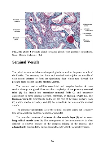
FIGURE  20.18  ■  Prostate  gland:  prostatic  glands  with  prostatic  concretions.
               Stain: Masson trichrome. ×64.


               Seminal Vesicle


               The paired seminal vesicles are elongated glands located on the posterior side of

               the bladder. The excretory duct from each seminal vesicle joins the ampulla of
               each  ductus  deferens  to  form  the  ejaculatory  duct,  which  runs  through  the
               prostate gland to open into the prostatic urethra.

                   The  seminal  vesicle  exhibits  convoluted  and  irregular  lumina.  A  cross
               section  through  the  gland  illustrates  the  complexity  of  the  primary  mucosal

               folds  (1)  that  branch  into  secondary  mucosal  folds  (2)  and  frequently
               anastomose  to  form  irregular  cavities,  chambers,  or  mucosal  crypts  (7).  The
               lamina propria (6) projects into and forms the core of the larger primary folds

               (1) and the smaller secondary folds (2) that extend into the lumen of the seminal
               vesicle.

                   The glandular epithelium (5) of the seminal vesicles varies but is usually
               low pseudostratified and low columnar or cuboidal.

                   The muscularis consists of an inner circular muscle layer (3) and an outer
               longitudinal muscle layer (4). This arrangement of the smooth muscles is often

               difficult  to  observe  because  of  the  complex  folding  of  the  mucosa.  The
               adventitia (8) surrounds the muscularis and blends with the connective tissue.


                                                          822