
Page 827 - Atlas of Histology with Functional Correlations
P. 827
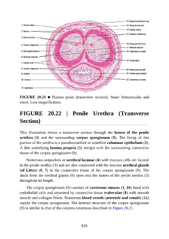
FIGURE  20.21  ■  Human  penis  (transverse  section).  Stain:  hematoxylin  and
               eosin. Low magnification.


               FIGURE  20.22  |  Penile  Urethra  (Transverse

               Section)


               This  illustration  shows  a  transverse  section  through  the  lumen  of  the  penile
               urethra  (3)  and  the  surrounding  corpus  spongiosum  (9).  The  lining  of  this
               portion of the urethra is a pseudostratified or stratified columnar epithelium (2).
               A thin underlying lamina propria (5) merges with the surrounding connective

               tissue of the corpus spongiosum (9).

                   Numerous outpockets or urethral lacunae (4) with mucous cells are located
               in the penile urethra (3) and are also connected with the mucous urethral glands
               (of Littre) (6,  7)  in  the  connective  tissue  of  the  corpus  spongiosum  (9).  The
               ducts from the urethral glands (6) open into the lumen of the penile urethra (3)

               throughout its length.

                   The corpus spongiosum (9) consists of cavernous sinuses (1, 10) lined with
               endothelial cells and separated by connective tissue trabeculae (8) with smooth
               muscle and collagen fibers. Numerous blood vessels (arteriole and venule) (11)

               supply the corpus spongiosum. The internal structure of the corpus spongiosum
               (9) is similar to that of the corpora cavernosa described in Figure 20.21.


                                                          826