
Page 824 - Atlas of Histology with Functional Correlations
P. 824
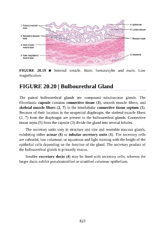
FIGURE  20.19  ■  Seminal  vesicle.  Stain:  hematoxylin  and  eosin.  Low
               magnification.


               FIGURE 20.20 | Bulbourethral Gland


               The  paired  bulbourethral  glands  are  compound  tubuloacinar  glands.  The

               fibroelastic capsule contains connective tissue (3), smooth muscle fibers, and
               skeletal muscle fibers (2, 7) in the interlobular connective tissue septum (5).
               Because of their location in the urogenital diaphragm, the skeletal muscle fibers
               (2, 7) from the diaphragm are present in the bulbourethral glands. Connective
               tissue septa (5) from the capsule (3) divide the gland into several lobules.


                   The secretory units vary in structure and size and resemble mucous glands,
               exhibiting either acinar (6) or tubular secretory units (1). The secretory cells
               are cuboidal, low columnar, or squamous and light staining with the height of the
               epithelial cells depending on the function of the gland. The secretory product of

               the bulbourethral glands is primarily mucus.

                   Smaller excretory ducts (4) may be lined with secretory cells, whereas the
               larger ducts exhibit pseudostratified or stratified columnar epithelium.


                                                          823