
Page 841 - Atlas of Histology with Functional Correlations
P. 841
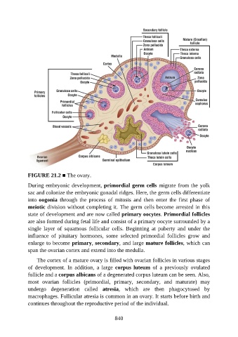
FIGURE 21.2 ■ The ovary.

               During embryonic development, primordial germ cells migrate from the yolk

               sac and colonize the embryonic gonadal ridges. Here, the germ cells differentiate
               into oogonia  through  the  process  of  mitosis  and  then  enter  the  first  phase  of
               meiotic division without completing it. The germ cells become arrested in this
               state of development and are now called primary oocytes. Primordial follicles
               are also formed during fetal life and consist of a primary oocyte surrounded by a

               single  layer  of  squamous  follicular  cells.  Beginning  at  puberty  and  under  the
               influence  of  pituitary  hormones,  some  selected  primordial  follicles  grow  and
               enlarge to become primary, secondary, and large mature follicles, which can

               span the ovarian cortex and extend into the medulla.

                   The cortex of a mature ovary is filled with ovarian follicles in various stages
               of  development.  In  addition,  a  large  corpus luteum  of  a  previously  ovulated
               follicle and a corpus albicans of a degenerated corpus luteum can be seen. Also,
               most  ovarian  follicles  (primordial,  primary,  secondary,  and  maturate)  may

               undergo  degeneration  called  atresia,  which  are  then  phagocytosed  by
               macrophages. Follicular atresia is common in an ovary. It starts before birth and
               continues throughout the reproductive period of the individual.


                                                          840