
Page 845 - Atlas of Histology with Functional Correlations
P. 845
Transformation of the postovulatory mature follicle into the corpus
luteum, a temporary endocrine organ.
Vascularization of the corpus luteum and, in response to LH, increased
production of progesterone and estrogen by the luteal cells.
Final maturation, or the second meiotic division of the secondary oocyte,
occurs at the time of fertilization by sperm. The liberated secondary oocyte
remains viable in the female reproductive tract for about 24 hours before it
begins to degenerate without completing the second meiotic division.
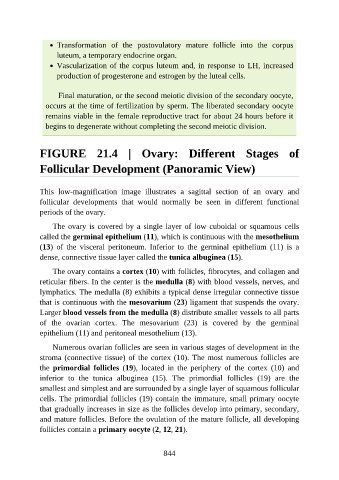
FIGURE 21.4 | Ovary: Different Stages of
Follicular Development (Panoramic View)
This low-magnification image illustrates a sagittal section of an ovary and
follicular developments that would normally be seen in different functional
periods of the ovary.
The ovary is covered by a single layer of low cuboidal or squamous cells
called the germinal epithelium (11), which is continuous with the mesothelium
(13) of the visceral peritoneum. Inferior to the germinal epithelium (11) is a
dense, connective tissue layer called the tunica albuginea (15).
The ovary contains a cortex (10) with follicles, fibrocytes, and collagen and
reticular fibers. In the center is the medulla (8) with blood vessels, nerves, and
lymphatics. The medulla (8) exhibits a typical dense irregular connective tissue
that is continuous with the mesovarium (23) ligament that suspends the ovary.
Larger blood vessels from the medulla (8) distribute smaller vessels to all parts
of the ovarian cortex. The mesovarium (23) is covered by the germinal
epithelium (11) and peritoneal mesothelium (13).
Numerous ovarian follicles are seen in various stages of development in the
stroma (connective tissue) of the cortex (10). The most numerous follicles are
the primordial follicles (19), located in the periphery of the cortex (10) and
inferior to the tunica albuginea (15). The primordial follicles (19) are the
smallest and simplest and are surrounded by a single layer of squamous follicular
cells. The primordial follicles (19) contain the immature, small primary oocyte
that gradually increases in size as the follicles develop into primary, secondary,
and mature follicles. Before the ovulation of the mature follicle, all developing
follicles contain a primary oocyte (2, 12, 21).
844