
Page 848 - Atlas of Histology with Functional Correlations
P. 848
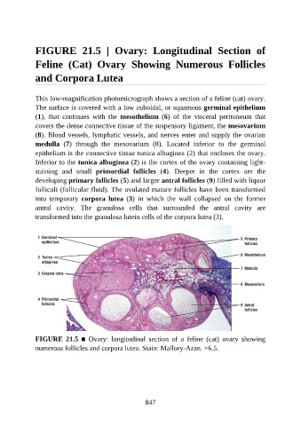
FIGURE  21.5  |  Ovary:  Longitudinal  Section  of

               Feline  (Cat)  Ovary  Showing  Numerous  Follicles

               and Corpora Lutea


               This low-magnification photomicrograph shows a section of a feline (cat) ovary.
               The surface is covered with a low cuboidal, or squamous germinal epithelium
               (1),  that  continues  with  the  mesothelium  (6)  of  the  visceral  peritoneum  that
               covers the dense connective tissue of the suspensory ligament, the mesovarium

               (8). Blood vessels, lymphatic vessels, and nerves enter and supply the ovarian
               medulla  (7)  through  the  mesovarium  (8).  Located  inferior  to  the  germinal
               epithelium is the connective tissue tunica albuginea (2) that encloses the ovary.
               Inferior to the tunica albuginea (2) is the cortex of the ovary containing light-

               staining  and  small  primordial  follicles  (4).  Deeper  in  the  cortex  are  the
               developing primary follicles (5) and larger antral follicles (9) filled with liquor
               folliculi (follicular fluid). The ovulated mature follicles have been transformed
               into  temporary  corpora  lutea  (3)  in  which  the  wall  collapsed  on  the  former

               antral  cavity.  The  granulosa  cells  that  surrounded  the  antral  cavity  are
               transformed into the granulosa lutein cells of the corpora lutea (3).


               FIGURE  21.5  ■  Ovary:  longitudinal  section  of  a  feline  (cat)  ovary  showing
               numerous follicles and corpora lutea. Stain: Mallory-Azan. ×6.5.


                                                          847