
Page 853 - Atlas of Histology with Functional Correlations
P. 853
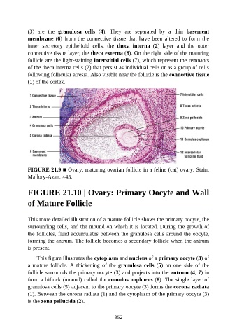
(3)  are  the  granulosa  cells  (4).  They  are  separated  by  a  thin  basement

               membrane (6)  from  the  connective  tissue  that  have  been  altered  to  form  the
               inner  secretory  epithelioid  cells,  the  theca  interna  (2)  layer  and  the  outer
               connective tissue layer, the theca externa (8). On the right side of the maturing
               follicle are the light-staining interstitial cells (7), which represent the remnants
               of the theca interna cells (2) that persist as individual cells or as a group of cells

               following follicular atresia. Also visible near the follicle is the connective tissue
               (1) of the cortex.


               FIGURE 21.9 ■ Ovary: maturing ovarian follicle in a feline (cat) ovary. Stain:

               Mallory-Azan. ×45.


               FIGURE 21.10 | Ovary: Primary Oocyte and Wall


               of Mature Follicle


               This more detailed illustration of a mature follicle shows the primary oocyte, the
               surrounding cells, and the mound on which it is located. During the growth of
               the follicles, fluid accumulates between the granulosa cells around the oocyte,

               forming the antrum. The follicle becomes a secondary follicle when the antrum
               is present.

                   This figure illustrates the cytoplasm and nucleus of a primary oocyte (3) of
               a  mature  follicle.  A  thickening  of  the  granulosa cells  (5)  on  one  side  of  the

               follicle surrounds the primary oocyte (3) and projects into the antrum (4, 7) in
               form a hillock (mound) called the cumulus oophorus (8). The single layer of
               granulosa cells (5) adjacent to the primary oocyte (3) forms the corona radiata
               (1). Between the corona radiata (1) and the cytoplasm of the primary oocyte (3)

               is the zona pellucida (2).


                                                          852