
Page 854 - Atlas of Histology with Functional Correlations
P. 854
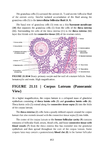
The granulosa cells (5) surround the antrum (4, 7) and secrete follicular fluid

               of  the  antrum  cavity.  Smaller  isolated  accumulation  of  the  fluid  among  the
               granulosa cells (5) is the intercellular follicular fluid (6, 9).

                   The basal row of granulosa cells (5) rests on a thin basement membrane
               (10) that separates the granulosa cells (5) from the cells of the theca interna
               (11). Surrounding the cells of the theca interna (11) is the theca externa (12)

               layer that blends with the connective tissue (13) of the ovarian cortex.


               FIGURE 21.10 ■ Ovary: primary oocyte and the wall of a mature follicle. Stain:
               hematoxylin and eosin. High magnification.


               FIGURE  21.11  |  Corpus  Luteum  (Panoramic


               View)


               At a higher magnification, the corpus luteum is a collapsed mass of glandular
               epithelium consisting of theca lutein cells (5) and granulosa lutein cells (6).

               Theca lutein cells (5) extend along the connective tissue septa (3) into the folds
               of the corpus luteum.

                   The theca externa (2) cells form a poorly defined capsule around the corpus
               luteum that also extends inward with the connective tissue septa (3) into folds.

                   The center of the corpus luteum or the former follicular cavity (9) contains

               remnants of follicular fluid, serum, blood cells, and loose connective tissue with
               blood vessels (7)  from  the  theca  externa  that  has  extended  into  the  glandular
               epithelium  and  then  spread  throughout  the  core  of  the  corpus  luteum.  Some
               corpora lutea may contain a postovulatory blood clot (8) in the former follicular


                                                          853