
Page 852 - Atlas of Histology with Functional Correlations
P. 852
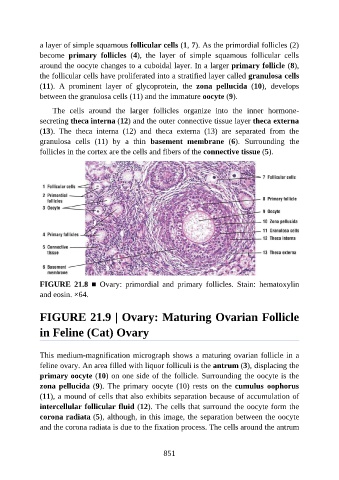
a layer of simple squamous follicular cells (1, 7). As the primordial follicles (2)

               become  primary  follicles  (4),  the  layer  of  simple  squamous  follicular  cells
               around the oocyte changes to a cuboidal layer. In a larger primary follicle (8),
               the follicular cells have proliferated into a stratified layer called granulosa cells
               (11).  A  prominent  layer  of  glycoprotein,  the  zona  pellucida  (10),  develops
               between the granulosa cells (11) and the immature oocyte (9).


                   The  cells  around  the  larger  follicles  organize  into  the  inner  hormone-
               secreting theca interna (12) and the outer connective tissue layer theca externa
               (13).  The  theca  interna  (12)  and  theca  externa  (13)  are  separated  from  the
               granulosa  cells  (11)  by  a  thin  basement  membrane  (6).  Surrounding  the

               follicles in the cortex are the cells and fibers of the connective tissue (5).


               FIGURE  21.8  ■  Ovary:  primordial  and  primary  follicles.  Stain:  hematoxylin
               and eosin. ×64.


               FIGURE 21.9 | Ovary: Maturing Ovarian Follicle

               in Feline (Cat) Ovary


               This medium-magnification micrograph shows a maturing ovarian follicle in a

               feline ovary. An area filled with liquor folliculi is the antrum (3), displacing the
               primary oocyte (10) on one side of the follicle. Surrounding the oocyte is the
               zona pellucida (9).  The  primary  oocyte  (10)  rests  on  the cumulus  oophorus
               (11), a mound of cells that also exhibits separation because of accumulation of
               intercellular follicular fluid (12). The cells that surround the oocyte form the

               corona radiata (5), although, in this image, the separation between the oocyte
               and the corona radiata is due to the fixation process. The cells around the antrum


                                                          851