
Page 850 - Atlas of Histology with Functional Correlations
P. 850
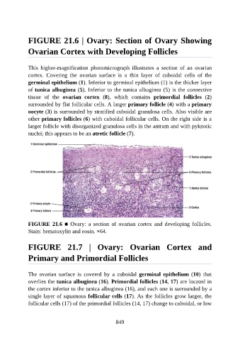
FIGURE 21.6 | Ovary: Section of Ovary Showing

               Ovarian Cortex with Developing Follicles


               This  higher-magnification  photomicrograph  illustrates  a  section  of  an  ovarian
               cortex.  Covering  the  ovarian  surface  is  a  thin  layer  of  cuboidal  cells  of  the

               germinal epithelium (1). Inferior to germinal epithelium (1) is the thicker layer
               of tunica albuginea (5). Inferior to the tunica albuginea (5) is the connective
               tissue  of  the  ovarian  cortex  (8),  which  contains  primordial  follicles  (2)

               surrounded by flat follicular cells. A larger primary follicle (4) with a primary
               oocyte (3) is surrounded by stratified cuboidal granulosa cells. Also visible are
               other primary follicles (6) with cuboidal follicular cells. On the right side is a
               larger follicle with disorganized granulosa cells in the antrum and with pyknotic
               nuclei; this appears to be an atretic follicle (7).


               FIGURE  21.6  ■  Ovary:  a  section  of  ovarian  cortex  and  developing  follicles.

               Stain: hematoxylin and eosin. ×64.


               FIGURE  21.7  |  Ovary:  Ovarian  Cortex  and


               Primary and Primordial Follicles


               The  ovarian  surface  is  covered  by  a  cuboidal  germinal  epithelium  (10)  that
               overlies the tunica albuginea (16). Primordial follicles (14, 17) are located in
               the cortex inferior to the tunica albuginea (16), and each one is surrounded by a

               single layer of squamous follicular cells (17). As the follicles grow larger, the
               follicular cells (17) of the primordial follicles (14, 17) change to cuboidal, or low


                                                          849