
Page 855 - Atlas of Histology with Functional Correlations
P. 855
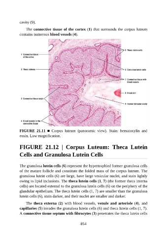
cavity (9).

                   The connective tissue of the cortex (1)  that  surrounds  the  corpus  luteum

               contains numerous blood vessels (4).


               FIGURE  21.11  ■  Corpus  luteum  (panoramic  view).  Stain:  hematoxylin  and

               eosin. Low magnification.


               FIGURE  21.12  |  Corpus  Luteum:  Theca  Lutein


               Cells and Granulosa Lutein Cells


               The granulosa lutein cells (6) represent the hypertrophied former granulosa cells
               of the mature follicle and constitute the folded mass of the corpus luteum. The

               granulosa lutein cells (6) are large, have large vesicular nuclei, and stain lightly
               owing to lipid inclusions. The theca lutein cells (1, 7) (the former theca interna
               cells) are located external to the granulosa lutein cells (6) on the periphery of the
               glandular epithelium. The theca lutein cells (1, 7) are smaller than the granulosa
               lutein cells (6), stain darker, and their nuclei are smaller and darker.


                   The theca externa  (2)  with  blood  vessels,  venule  and  arteriole  (4),  and
               capillaries (5) invades the granulosa lutein cells (6) and theca lutein cells (1, 7).
               A connective tissue septum with fibrocytes (3) penetrates the theca lutein cells


                                                          854