
Page 860 - Atlas of Histology with Functional Correlations
P. 860
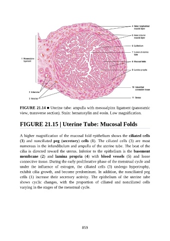
FIGURE 21.14 ■ Uterine tube: ampulla with mesosalpinx ligament (panoramic

               view, transverse section). Stain: hematoxylin and eosin. Low magnification.


               FIGURE 21.15 | Uterine Tube: Mucosal Folds


               A higher magnification of the mucosal fold epithelium shows the ciliated cells
               (3)  and  nonciliated  peg  (secretory)  cells  (1).  The  ciliated  cells  (3)  are  most
               numerous in the infundibulum and ampulla of the uterine tube. The beat of the
               cilia  is  directed  toward  the  uterus.  Inferior  to  the  epithelium  is  the  basement

               membrane  (2)  and  lamina  propria  (4)  with  blood  vessels  (5)  and  loose
               connective tissue. During the early proliferative phase of the menstrual cycle and
               under  the  influence  of  estrogen,  the  ciliated  cells  (3)  undergo  hypertrophy,
               exhibit cilia growth, and become predominant. In addition, the nonciliated peg

               cells  (1)  increase  their  secretory  activity.  The  epithelium  of  the  uterine  tube
               shows  cyclic  changes,  with  the  proportion  of  ciliated  and  nonciliated  cells
               varying in the stages of the menstrual cycle.


                                                          859