
Page 863 - Atlas of Histology with Functional Correlations
P. 863
maximum during the follicular phase, at which time the ovarian follicles are
maturing and circulating levels of estrogen are high.
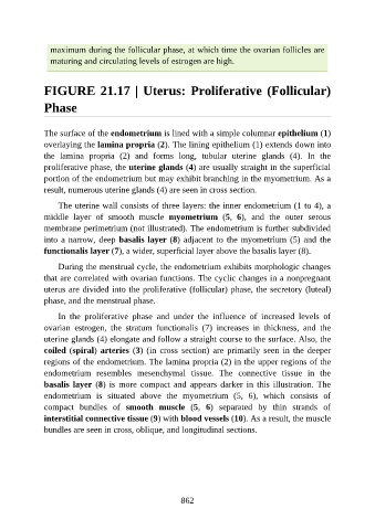
FIGURE 21.17 | Uterus: Proliferative (Follicular)
Phase
The surface of the endometrium is lined with a simple columnar epithelium (1)
overlaying the lamina propria (2). The lining epithelium (1) extends down into
the lamina propria (2) and forms long, tubular uterine glands (4). In the
proliferative phase, the uterine glands (4) are usually straight in the superficial
portion of the endometrium but may exhibit branching in the myometrium. As a
result, numerous uterine glands (4) are seen in cross section.
The uterine wall consists of three layers: the inner endometrium (1 to 4), a
middle layer of smooth muscle myometrium (5, 6), and the outer serous
membrane perimetrium (not illustrated). The endometrium is further subdivided
into a narrow, deep basalis layer (8) adjacent to the myometrium (5) and the
functionalis layer (7), a wider, superficial layer above the basalis layer (8).
During the menstrual cycle, the endometrium exhibits morphologic changes
that are correlated with ovarian functions. The cyclic changes in a nonpregnant
uterus are divided into the proliferative (follicular) phase, the secretory (luteal)
phase, and the menstrual phase.
In the proliferative phase and under the influence of increased levels of
ovarian estrogen, the stratum functionalis (7) increases in thickness, and the
uterine glands (4) elongate and follow a straight course to the surface. Also, the
coiled (spiral) arteries (3) (in cross section) are primarily seen in the deeper
regions of the endometrium. The lamina propria (2) in the upper regions of the
endometrium resembles mesenchymal tissue. The connective tissue in the
basalis layer (8) is more compact and appears darker in this illustration. The
endometrium is situated above the myometrium (5, 6), which consists of
compact bundles of smooth muscle (5, 6) separated by thin strands of
interstitial connective tissue (9) with blood vessels (10). As a result, the muscle
bundles are seen in cross, oblique, and longitudinal sections.
862