
Page 861 - Atlas of Histology with Functional Correlations
P. 861
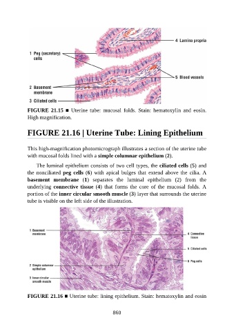
FIGURE 21.15 ■  Uterine  tube:  mucosal  folds.  Stain:  hematoxylin  and  eosin.

               High magnification.


               FIGURE 21.16 | Uterine Tube: Lining Epithelium


               This high-magnification photomicrograph illustrates a section of the uterine tube
               with mucosal folds lined with a simple columnar epithelium (2).

                   The luminal epithelium consists of two cell types, the ciliated cells (5) and
               the nonciliated peg cells (6) with apical bulges that extend above the cilia. A
               basement  membrane  (1)  separates  the  luminal  epithelium  (2)  from  the

               underlying connective tissue (4)  that  forms  the  core  of  the  mucosal  folds.  A
               portion of the inner circular smooth muscle (3) layer that surrounds the uterine
               tube is visible on the left side of the illustration.


               FIGURE 21.16 ■ Uterine tube: lining epithelium. Stain: hematoxylin and eosin


                                                          860