
Page 857 - Atlas of Histology with Functional Correlations
P. 857
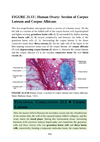
FIGURE 21.13 | Human Ovary: Section of Corpus

               Luteum and Corpus Albicans


               This low-magnification micrograph shows a section of a human ovary. On the
               left side is a section of the folded wall of the corpus luteum with hypertrophied

               and lighter-staining granulosa lutein cells (3, 5) surrounded by darker-staining
               theca  lutein  cells  (1,  4)  located  peripherally  and  between  the  folds  of  the
               granulosa  lutein  cells  (3,  5).  Surrounding  the  corpus  luteum  is  the  dense

               connective tissue layer theca externa (2). On the right side of the figure is the
               blue-staining connective tissue scar of the corpus luteum, the corpus albicans
               (7) with degenerating corpus luteum (6) above it. Between the corpus luteum
               and  the  corpus  albicans  (7)  is  the  vascular  connective  tissue  (9)  with  blood
               vessels (8).


               FIGURE 21.13 ■ Human ovary: a section of corpus luteum and corpus albicans.
               Stain: Mallory-Azan. ×10.5.


                  FUNCTIONAL  CORRELATIONS  21.2  ■  Corpus


                  Luteum


                 After the mature follicle liberates the secondary oocyte into the infundibulum
                 of the uterine tube, the wall of the ruptured mature follicle collapses, and the

                 ovary  enters  the  luteal  phase.  During  this  luteinization  phase,  luteinizing
                 hormone (LH) secretion induces hypertrophy and transforms the granulosa
                 cells  and  theca  interna  cells  into  granulosa  lutein  cells  and  theca  lutein
                 cells, respectively, forming a temporary endocrine tissue, the corpus luteum.


                                                          856