
Page 851 - Atlas of Histology with Functional Correlations
P. 851
columnar, and the follicles now become primary follicles (4, 11). The
developing oocytes (4, 13) in the follicles also have a large eccentric nucleus (7,
13) with a nucleolus.
In primary (growing) follicles (4, 11), the follicular cells proliferate by
mitosis (3) and form layers of cuboidal cells called the granulosa cells (8, 12)
that surround the primary oocytes (4, 13). A single layer of the granulosa cells
around the oocyte forms the corona radiata (5).
Between the corona radiata (5) and the oocyte appears the noncellular
glycoprotein layer called the zona pellucida (6). The stromal cells that surround
the follicular cells now differentiate into the theca interna (9) layer located
adjacent to the granulosa cells (8, 12). A thin basement membrane (not shown)
separates the granulosa cells (8, 12) from the theca interna (9) cells.
Many primordial, developing, or mature follicles exhibit degeneration, die,
and are lost through atresia. A degenerating atretic follicle (1) is illustrated in
the upper left corner of the illustration. Numerous blood vessels (2) surround the
developing follicles in the connective tissue of the cortex (15).
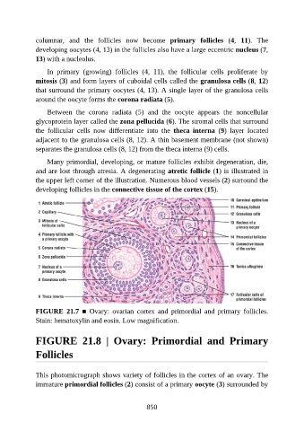
FIGURE 21.7 ■ Ovary: ovarian cortex and primordial and primary follicles.
Stain: hematoxylin and eosin. Low magnification.
FIGURE 21.8 | Ovary: Primordial and Primary
Follicles
This photomicrograph shows variety of follicles in the cortex of an ovary. The
immature primordial follicles (2) consist of a primary oocyte (3) surrounded by
850