
Page 913 - Atlas of Histology with Functional Correlations
P. 913
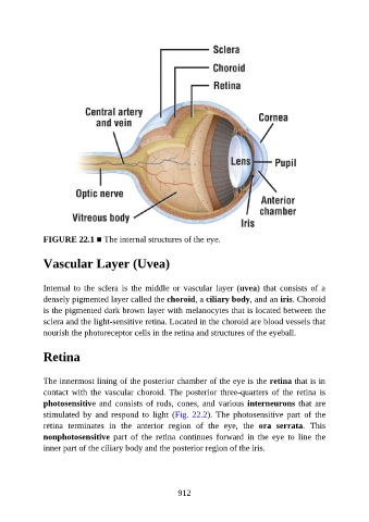
FIGURE 22.1 ■ The internal structures of the eye.


               Vascular Layer (Uvea)


               Internal  to  the  sclera  is  the  middle  or  vascular  layer  (uvea)  that  consists  of  a

               densely pigmented layer called the choroid, a ciliary body, and an iris. Choroid
               is the pigmented dark brown layer with melanocytes that is located between the
               sclera and the light-sensitive retina. Located in the choroid are blood vessels that
               nourish the photoreceptor cells in the retina and structures of the eyeball.


               Retina


               The innermost lining of the posterior chamber of the eye is the retina that is in
               contact with the vascular choroid. The posterior three-quarters of the retina is

               photosensitive and consists of rods, cones, and various interneurons that are
               stimulated  by  and  respond  to  light  (Fig. 22.2).  The  photosensitive  part  of  the
               retina  terminates  in  the  anterior  region  of  the  eye,  the  ora  serrata.  This
               nonphotosensitive  part  of  the  retina  continues  forward  in  the  eye  to  line  the

               inner part of the ciliary body and the posterior region of the iris.


                                                          912