
Page 918 - Atlas of Histology with Functional Correlations
P. 918
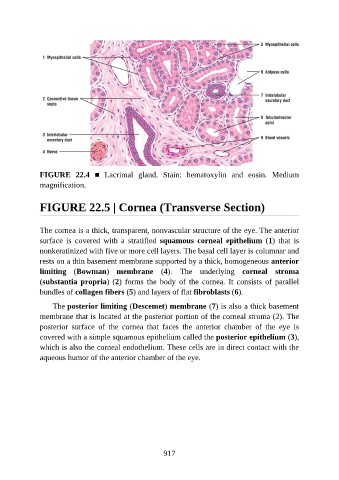
FIGURE  22.4  ■  Lacrimal  gland.  Stain:  hematoxylin  and  eosin.  Medium
               magnification.


               FIGURE 22.5 | Cornea (Transverse Section)


               The cornea is a thick, transparent, nonvascular structure of the eye. The anterior
               surface  is  covered  with  a  stratified  squamous  corneal  epithelium  (1)  that  is
               nonkeratinized with five or more cell layers. The basal cell layer is columnar and

               rests on a thin basement membrane supported by a thick, homogeneous anterior
               limiting  (Bowman)  membrane  (4).  The  underlying  corneal  stroma
               (substantia propria) (2)  forms  the  body  of  the  cornea.  It  consists  of  parallel
               bundles of collagen fibers (5) and layers of flat fibroblasts (6).

                   The posterior limiting (Descemet) membrane (7) is also a thick basement

               membrane that is located at the posterior portion of the corneal stroma (2). The
               posterior  surface  of  the  cornea  that  faces  the  anterior  chamber  of  the  eye  is
               covered with a simple squamous epithelium called the posterior epithelium (3),

               which is also the corneal endothelium. These cells are in direct contact with the
               aqueous humor of the anterior chamber of the eye.


                                                          917