
Page 921 - Atlas of Histology with Functional Correlations
P. 921
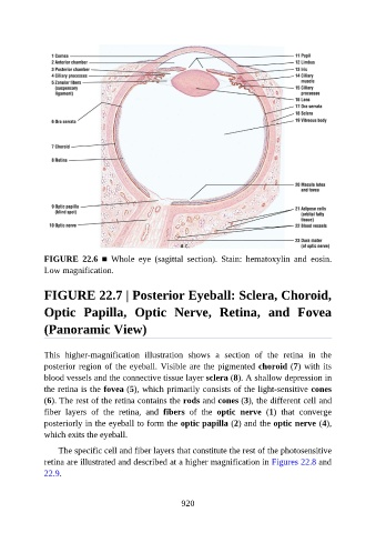
FIGURE  22.6  ■  Whole  eye  (sagittal  section).  Stain:  hematoxylin  and  eosin.

               Low magnification.


               FIGURE 22.7 | Posterior Eyeball: Sclera, Choroid,


               Optic  Papilla,  Optic  Nerve,  Retina,  and  Fovea

               (Panoramic View)


               This  higher-magnification  illustration  shows  a  section  of  the  retina  in  the

               posterior region of the eyeball. Visible are the pigmented choroid (7) with its
               blood vessels and the connective tissue layer sclera (8). A shallow depression in
               the retina is the fovea (5), which primarily consists of the light-sensitive cones
               (6). The rest of the retina contains the rods and cones (3), the different cell and

               fiber  layers  of  the  retina,  and  fibers  of  the  optic  nerve  (1)  that  converge
               posteriorly in the eyeball to form the optic papilla (2) and the optic nerve (4),
               which exits the eyeball.

                   The specific cell and fiber layers that constitute the rest of the photosensitive
               retina are illustrated and described at a higher magnification in Figures 22.8 and

               22.9.


                                                          920